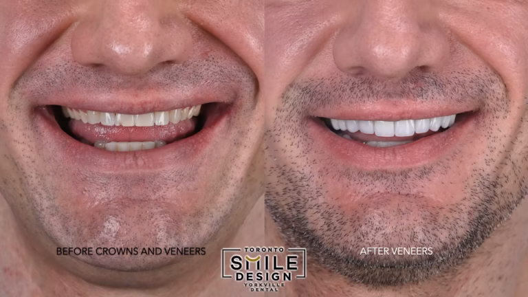
You might be here because someone mentioned a root canal, or your dentist showed you a dark shadow on an X-ray, and now your mind will not stop racing. You are wondering what is really going on inside that tooth, whether the treatment will work, and how your endodontist can be so sure about what you actually need. If you are considering endodontic treatment in Bolingbrook, understanding your options and the process can help ease your concerns and prepare you for your next appointment.
That worry makes sense. For years, people had to trust flat, blurry images and a lot of guesswork. Today, though, the future of endodontics with digital imaging is changing that story. In simple terms, modern imaging helps your endodontist see more, treat more precisely, and protect more of your natural tooth. You get clearer answers, more targeted care, and usually a smoother recovery.
So, where does that leave you? If you are anxious, the short version is this. New digital tools, especially 3D scans, give a far more accurate picture of your tooth and the bone around it. That means fewer surprises, better decisions, and often fewer procedures over the long run.
Why traditional X-rays sometimes leave you worried and confused
Think about the last time you had a dental X-ray. You bit down on a little tab, the assistant stepped out of the room, there was a quick buzz, and then your dentist stared at a small, gray image on a screen. You might have nodded, even though you could not really tell what you were looking at.
That is the first part of the problem. Standard 2D X-rays flatten a 3D tooth into a single slice. Important details can hide behind roots or bone, tiny fractures might be invisible, and the true shape of the root canals is hard to judge. Because of this, even a very skilled endodontist sometimes has to work with incomplete information.
When information is incomplete, your stress grows. You may be told that a tooth “might” need retreatment or that an infection “could” be present, and you are left choosing between waiting and acting, without feeling certain either way. Financially, that is tough too. No one wants to pay for a procedure and then discover later that something was missed.
So the tension builds. You want to save your tooth, avoid pain, and protect your budget, yet you do not feel you can really see what your doctor sees. That is exactly where digital imaging and advanced endodontic imaging start to change the experience.
How 3D digital imaging reshapes modern root canal care
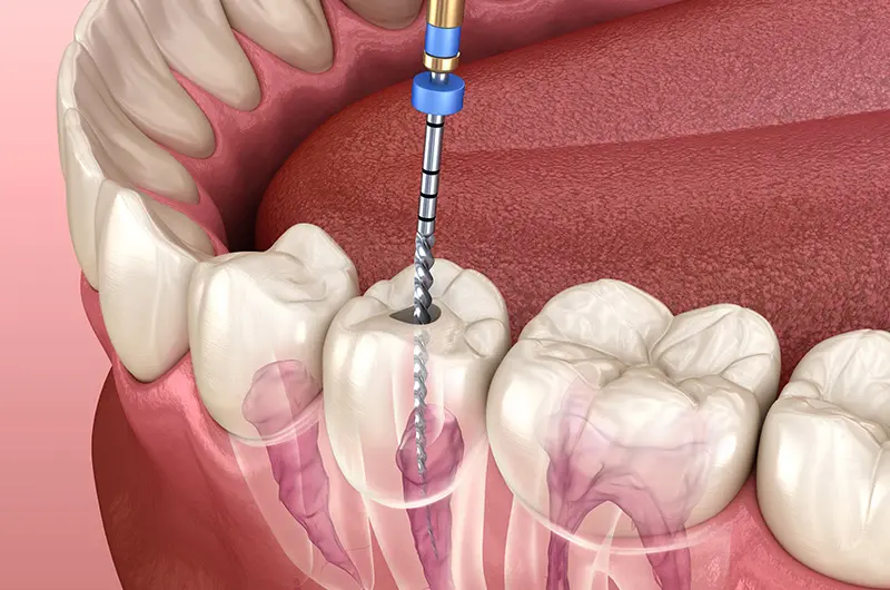
Digital imaging covers different tools, but the one that is reshaping root canal treatment the most is cone beam computed tomography, often shortened to CBCT. Instead of a flat X-ray, CBCT takes a 3D scan of your teeth and jaws so your endodontist can look from different angles and slice through the image layer by layer.
The American Association of Endodontists has published detailed guidance on when and how CBCT should be used. If you are curious about the professional standards behind these scans, you can read their overview of cone beam computed tomography in endodontics.
So what does this mean for you in the chair? Imagine a few common situations.
- You have a stubborn toothache, but regular X-rays look “normal.” A 3D scan can reveal a hidden extra canal, a tiny crack, or a small area of bone loss that a 2D image missed.
- You had a root canal years ago, and the tooth is acting up again. A CBCT image can show whether there is a missed canal, a small fracture, or a persistent infection at the root tip.
- Your dentist is unsure if a dark area at the end of a root is a true infection or just an anatomical variation. The 3D view can make that distinction much clearer and help avoid unnecessary treatment.
Researchers are also studying how AI and advanced software can interpret these 3D images, improving the detection of fractures and lesions. For example, recent work published in medical databases such as PubMed on CBCT and diagnostic accuracy suggests that digital imaging can reveal problems that simple X-rays consistently miss.
Because of this, the future of endodontics with digital imaging is not just about fancy pictures. It is about more accurate diagnoses, targeted root canal procedures, and a better chance of saving teeth that might once have been extracted.
But what about radiation, cost, and safety
With any new technology, there are fair questions. You might wonder about radiation exposure or worry that you are being pushed toward an expensive scan you do not really need.
Here is the nuance. CBCT uses more radiation than a single small dental X-ray, but often less than a full set of conventional images taken from several angles. Modern machines also allow your endodontist to limit the scanned area to just the region of interest. Professional bodies have issued position statements on using CBCT carefully and only when the expected benefit is real. If you want the more technical version of those guidelines, you can look at the AAE’s formal cone beam computed tomography statement.
Financially, a 3D scan is an extra cost. The key question is whether it helps avoid repeat procedures, failed treatments, or unnecessary extractions. Often it does. A precise diagnosis can prevent you from spending time and money on treatments that are unlikely to succeed.
So, where does that leave you? It leaves you with a decision. You do not have to simply accept or reject digital imaging. You can ask clear questions, weigh the benefits against the risks and costs, and choose what feels right for your health and your budget.
Comparing traditional X-rays and 3D digital imaging in endodontics
To make things more concrete, here is a simple comparison of conventional 2D X-rays and CBCT-based digital endodontic imaging.
| Aspect | Traditional 2D Dental X-rays | CBCT 3D Digital Imaging |
|---|---|---|
| View of tooth and roots | Flat image. Structures can overlap and hide details. | Three-dimensional view. Roots, canals, and surrounding bone are seen from many angles. |
| Detection of extra canals | Often missed, especially in molars. | Much higher chance of finding additional or unusual canals. |
| Ability to see small fractures | Limited. Many cracks are not visible. | Better detection of vertical root fractures and fine cracks. |
| Radiation exposure | Low per image. Multiple images may be needed for diagnosis. | Higher per scan, but usually a single focused scan replaces several images. |
| Usefulness in complex cases | Can leave questions unanswered or require guesswork. | Clarifies anatomy and pathology, especially in retreatment or surgical cases. |
| Impact on treatment planning | Good for straightforward cases. | Stronger support for accurate planning in complicated or uncertain cases. |
Three practical steps you can take before your next endodontic visit
You do not need to become a radiology expert. You just need a clear way to talk with your endodontist and to understand your options.
1. Ask, “What are you hoping to see with this image?”
Before any X-ray or 3D scan, ask what specific question your endodontist is trying to answer. For example, “Are you checking for a hidden canal, a fracture, or an infection at the tip of the root?” When you know the purpose, it is easier to judge whether the scan feels worthwhile.
2. Ask for a short tour of your images
When your images are ready, ask your endodontist to walk you through them in plain language. You can say, “Can you show me where the problem is and how this imaging changed your treatment plan?” Seeing the difference between a 2D and 3D image on the screen can reduce your anxiety and help you feel more in control.
3. Weigh cost and benefit openly
If cost or radiation exposure worries you, say so. Ask if a CBCT scan is truly necessary or just “nice to have” in your case. A thoughtful endodontist will explain whether traditional imaging is enough or why the extra information from a scan might prevent repeat procedures or an extraction later. You can then decide with a clearer head instead of under pressure.
Looking ahead with more clarity and less fear
Facing root canal treatment or ongoing tooth pain is never easy. It touches your comfort, your wallet, and your sense of control over your own body. You are allowed to feel uneasy about that.
The encouraging news is that as future-focused endodontic care continues to embrace digital imaging, your choices become better informed. You are no longer asked to trust a blurry shadow and a shrug. You can see more, understand more, and decide from a place of knowledge rather than fear.
The next step is simple. At your consultation, bring your questions about imaging and ask how these tools could affect your diagnosis and treatment. A clear, calm conversation is often the best pain relief you can get before any procedure even begins.